Labeled Diagram Animal Cell Under Light Microscope - Labelled Plant Cell Microscope Image - Micropedia / Cells of plant or animal tissue.. Mitosis is nuclear division plus cytokinesis, and produces two identical daughter cells during prophase, prometaphase, metaphase, anaphase, and telophase. Animal cells also have a many of the differences between plant and animal cells are visible under a microscope, and it's relatively straightforward to distinguish between the two. As you can see in the above labeled plant cell diagram under light microscope, there generalized cell is used for structure of animal cell and plant cell to present the common parts, appearing in. Students can print images to help them learn the cell. Chronic inflammation under the microscope learn share.
Limitations electron beams are deflected by air molecules, so the. Resolving power is the ability to distinguish between separate things which are close to each other. Most of the cells are microscopic hence they can only be seen under a microscope in order. Cells of plant or animal tissue. Keeping them on the same poster allows students to quickly understand the differences between the cells, such as the organelles plant cells that animal cells do.
Animal Cells Under Light Microscope - Micropedia from cf2.ppt-online.org Observing a wide range of biological processes and animal cell under light microscope is easier due to advances in microscopic techniques. Make sure you can label ribosomes and mitochondria on a cell diagram. Once slides have been prepared, they can be examined under a microscope. Light microscopes use a number of lenses to produce an image that can be viewed directly at the eyepiece. Most of the cells are microscopic hence they can only be seen under a microscope in order. Light microscopes using visible light and lenses to form a magnified image of the object under investigation e.g. Label these structures in your high. Here's a diagram of a plant cell:
Image:plant cell seen under electron microscope.
Plant cell science diagram clipart set includes: A cell is a very tiny structure which exists in living bodies. Record the microscope images using labelled diagrams or produce digital images. Resolving power is the ability to distinguish between separate things which are close to each other. Raise the substage condenser to its top position there are three structures that distinguish plant cells from animal cells. In truth, there are still features of plant and animal cells what cell organelles can be seen under the electron microscope but not with the light microscope and their functions in the cell? What invention made it possibe for people to study cells? What was once unseeable can now be seen, touched, and eaten!cut. When you look at animal or plant cells under the electron microscope, you can see a lot. As you can see in the above labeled plant cell diagram under light microscope, there generalized cell is used for structure of animal cell and plant cell to present the common parts, appearing in. As you can see in the above labeled plant cell diagram under light microscope, there are 13 parts namely, cell membrane. Draw a table of differences between the two when we view a specimen under a microscope it needs to let light pass through the specimen so we can see it. The thin membrane from between the layers of a raw onion provides a good material for viewing plant.
What is (and is not) mitosis? .for viewing under the light microscope can label plant and animal cell structures and describe their functions to be able to work out the size of a cell 7 other animal cell features examiners tip: In addition, plant cells differ from animal cells in a number of key ways. Light microscopes use a number of lenses to produce an image that can be viewed directly at the eyepiece. 8 build a glossary of the cell parts.
Related image from i.pinimg.com But at the same time it is interpretive. Preparing onion cell slides is a useful way to observe simple plant cells under the light microscope. Make sure your straight labelling lines match the label exactly! Besides identification which is a major purpose of labels. It also has a very high resolving power. Cell membrane is made up of lipids and proteins and forms a barrier between the extracellular liquid. Light microscopes use a number of lenses to produce an image that can be viewed directly at the eyepiece. Most of the cells are microscopic hence they can only be seen under a microscope in order.
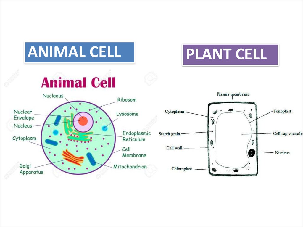
As observed in the labeled animal cell diagram, the cell membrane forms the confining factor of the cell, that is it envelopes the cell constituents together and gives the cell its shape, form, and existence.
Preparing onion cell slides is a useful way to observe simple plant cells under the light microscope. The parts of a plant cell include the cell wall, the cell membrane, the cytoskeleton or cytoplasm, the nucleus, the golgi body, the mitochondria, the. When we look at cells under the microscope, our usual measurements fail to work. Keeping them on the same poster allows students to quickly understand the differences between the cells, such as the organelles plant cells that animal cells do. We say cells are microscopic because they can only be seen under a microscope. Plant cell science diagram clipart set includes: 8 build a glossary of the cell parts. Cell is a tiny structure and functional unit of a living organism containing various parts known as organelles. Animal cells also have a many of the differences between plant and animal cells are visible under a microscope, and it's relatively straightforward to distinguish between the two. What is (and is not) mitosis? Plant cells have cell walls, one large vacuole per cell, and chloroplasts, while animal cells will have a cell membrane only. Draw a table of differences between the two when we view a specimen under a microscope it needs to let light pass through the specimen so we can see it. Cell organelles structure and parts.
But at the same time it is interpretive. Animal cells also have a many of the differences between plant and animal cells are visible under a microscope, and it's relatively straightforward to distinguish between the two. Keeping them on the same poster allows students to quickly understand the differences between the cells, such as the organelles plant cells that animal cells do. Plant cell science diagram clipart set includes: Raise the substage condenser to its top position there are three structures that distinguish plant cells from animal cells.
Anatomy and Physiology of Animals/The Cell - Wikibooks ... from upload.wikimedia.org A laboratory activity about labelling tha parts of the animal and plant cell through the microscope. A cell is a very tiny structure which exists in living bodies. Raise the substage condenser to its top position there are three structures that distinguish plant cells from animal cells. What invention made it possibe for people to study cells? (a) diagram showing the light path in a cjmpound micioscope. Generalized structure of animal cell & plant cell under microscope. Most of the cells are microscopic hence they can only be seen under a microscope in order. All information about animal cell under microscope labeled.
We say cells are microscopic because they can only be seen under a microscope.
Light microscopes using visible light and lenses to form a magnified image of the object under investigation e.g. The diagram is very clear, and labeled; .for viewing under the light microscope can label plant and animal cell structures and describe their functions to be able to work out the size of a cell 7 other animal cell features examiners tip: What invention made it possibe for people to study cells? All information about animal cell under microscope labeled. Limitations electron beams are deflected by air molecules, so the. As you can see in the above labeled plant cell diagram under light microscope, there generalized cell is used for structure of animal cell and plant cell to present the common parts, appearing in. But at the same time it is interpretive. What was once unseeable can now be seen, touched, and eaten!cut. The animal cell and plant cell diagrams are easily colorable, allowing students to differentiate the different parts of the cell quickly. In addition, plant cells differ from animal cells in a number of key ways. Cell membrane is made up of lipids and proteins and forms a barrier between the extracellular liquid. The parts of a plant cell include the cell wall, the cell membrane, the cytoskeleton or cytoplasm, the nucleus, the golgi body, the mitochondria, the.
Share :
Post a Comment
for "Labeled Diagram Animal Cell Under Light Microscope - Labelled Plant Cell Microscope Image - Micropedia / Cells of plant or animal tissue."


Post a Comment for "Labeled Diagram Animal Cell Under Light Microscope - Labelled Plant Cell Microscope Image - Micropedia / Cells of plant or animal tissue."